In this page you will find a set of 3D meshes of a true human brain, which were constructed from 12 volumes acquired using magnetic resonance imaging (mri). These meshes can be used in a variety of compositions, for both scientific or artistic purposes.
Image acquisition and reconstruction
The images were acquired at the Research Imaging Institute, University of Texas Health Science Center at San Antonio, in a Siemens magnetom Trio 3T system, in two sessions, each consisting of 6 acquisitions of T1-weighted images, using a mprage sequence, with voxel size of 0.8×0.8×0.8 milimeters. The images were registered and averaged to improve signal-to-noise ratio, as described here, and bias corrected using spm8 software. The already realigned, averaged and bias-corrected volume, in nifti format, is available here.
The generation of the cortical meshes and subcortical segmentations used FreeSurfer 5.2.0. The splitting of the cortical meshes into independent objects was performed using a custom script that soon will be released at Brainder.org (update: they are now available here). The subcortical meshes were produced from the volumetric segmentations, as described here.
File formats and uses
 The files below are provided in the ascii versions of three different formats: FreeSurfer surface (*.srf), Chris Rorden’s highly efficient MZ3 format (*.mz3), Wavefront Object (*.obj) and Stanford Polygon (*.ply). Click here for more information on these formats; for mz3, click here.
Although the title suggests that these files are meant to be used with Blender, they can in fact be used with any computer graphics application that can import these formats. This includes 3D Studio max, daz Studio, Houdini, Autodesk Maya, FreeCAD, Rhino, MeshLab, and many others.
Cortical meshes
Note that the small figures below were generated using the FreeSurfer utility tksurfer, but the actual model files do not contain any colours. They should be coloured or textured using your own creativity, in your favourite modelling software.
| Pial | White | Inflated | |
|---|---|---|---|
| Full hemispheres, each as a single mesh. |
 |
 |
 |
srf mz3 obj ply |
srf mz3 obj ply |
srf mz3 obj ply |
|
| Cortical mesh split according to the Desikan-Killiany atlas, one region per file. |
 |
 |
 |
srf mz3 obj ply |
srf mz3 obj ply |
srf mz3 obj ply |
|
| Cortical mesh split according to the Destrieux et al. atlas, one region per file. |
 |
 |
 |
srf mz3 obj ply |
srf mz3 obj ply |
srf mz3 obj ply |
|
| Cortical mesh split according to the Desikan-Killiany-Tourville (DKT) atlas, one region per file. |
 |
 |
 |
srf mz3 obj ply |
srf mz3 obj ply |
srf mz3 obj ply |
Subcortical structures
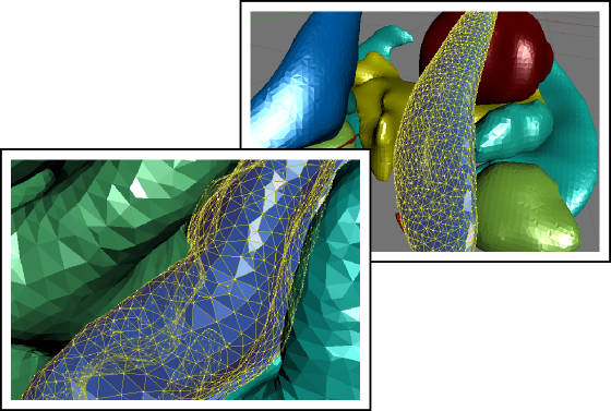
In addition to the above cortical meshes, surfaces for subcortical structures are also available. These are not produced directly by the FreeSurfer pipeline. However, the segmented volumes that are part of the subcortical stream can be used to generate surfaces for visualisation purposes, as described here. The meshes for the same brain, in different formats, can be downloaded here: srf mz3 obj ply.
Download all meshes in a single file
To download all the models at once, choose the format you want here: srf mz3 obj ply.
References
The details of each of the atlases above are in the following publications:
- Desikan RS, Ségonne F, Fischl B, Quinn BT, Dickerson BC, Blacker D, Buckner RL, Dale AM, Maguire RP, Hyman BT, Albert MS, Killiany RJ. An automated labeling system for subdividing the human cerebral cortex on MRI scans into gyral based regions of interest. Neuroimage. 2006 Jul 1;31(3):968-80.
- Destrieux C, Fischl B, Dale A, Halgren E. Automatic parcellation of human cortical gyri and sulci using standard anatomical nomenclature. Neuroimage. 2010 Oct 15;53(1):1-15.
- Klein A, Tourville J. 101 labeled brain images and a consistent human cortical labeling protocol. Front Neurosci. 2012;6:171.
License
The models in this page are by Anderson Winkler and are licensed under a Creative Commons Attribution-ShareAlike 3.0 Unported License. The original work can be found at https://brainder.org/brain-for-blender.

